Biological and abiotic factors influence eDNA Degradation in aquatic systems
Article 1:
Title:Water Flow and Biofilm Cover Influence Environmental DNA Detection in Recirculating Streams
Download website: https://doi.org/10.1021/acs.est.8b01822
Abstract : The increasing use of environmental DNA(eDNA) for determination of species presence in aquatic ecosystems is an invaluable technique for both ecology as a field and for the management of aquatic ecosystems. We examined the degradation dynamics of fish eDNA using an experimental array of recirculating streams, also using a “nested” primer assay to estimate degradation among eDNA fragment sizes. We introduced eDNA into streams with a range of water velocities (0.1−0.8 m s−1) and substrate biofilm coverage (0−100%) and monitored eDNA concentrations over time (∼10 d) to assess how biophysical conditions influence eDNA persistence. We found that the presence of biofilm significantly increased initial decay rates relative to previous studies conducted in nonflowing microcosms, suggesting important differences in detection and persistence in lentic vs lotic systems. Lastly, by using a nested primer assay that targeted different size eDNA fragments, we found that fragment size altered both the estimated rate constant coefficients, as well as eDNA detectability over time. Larger fragments (>600 bp) were quickly degraded, while shorter fragments (<100 bp) remained detectable for the entirety of the experiment. When using eDNA as a stream monitoring tool, understanding environmental factors controlling eDNA degradation will be critical for optimizing eDNA sampling strategies.
Main content:
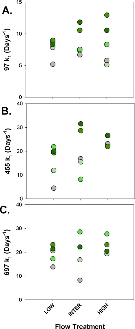
Figure 1. Estimated primary (k1) degradation terms from biphasicmodel fitting for A) 97 bp, B) 455 bp, and C) 697 bp eDNAfragments across all velocity treatments (x-axis). Color gradient ofdots represents biofilm cover treatment, from gray (0%) to dark green(100%). Secondary degradation terms are reported in SI Table 2.
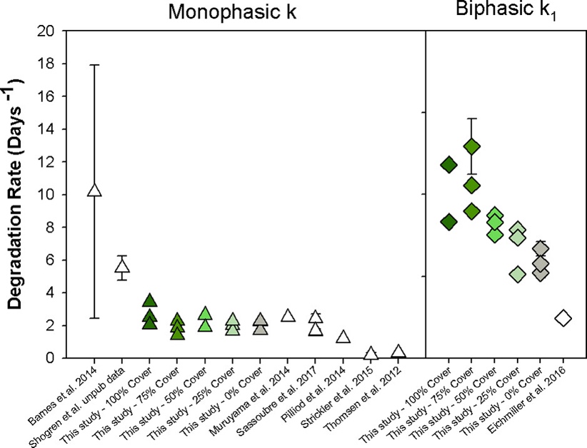
Figure 2. Meta-analysis of previously published studies on fish eDNArate constant coefficients (white) and this study (gray to greengradient) for monophasic (triangles) and biphasic (diamond) rateconstants (k vs k1). Rate constants expressed in days−1 (±SE ifreported).
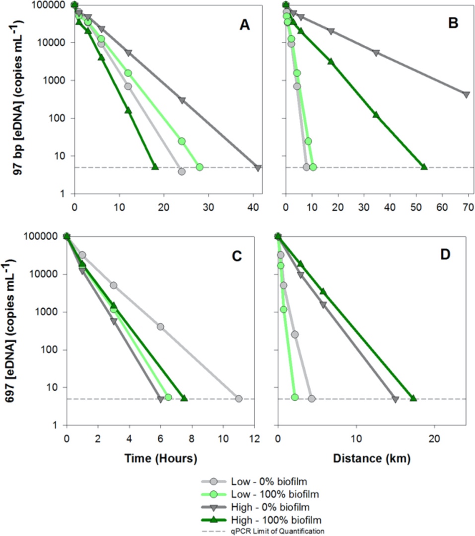
Figure 3. Projected (i.e., modeled) eDNA concentration in time (A: 97 bp fragment, C: 697 bp fragment) and distance (B: 97 bp fragment, D: 697bp fragment) based on 4 flow/biofilm scenarios (Low-0%, Low-100%, High-0%, and High-100% biofilm coverage) using the estimated k1 termfrom the artificial stream experiment.
Conclusion:
Biphasic Degradation Is an Important “Fate” of eDNA.
Flow Had Little Effect on Rate Constants but Increased Potential eDNA Transport Distances.
Biofilm Increased eDNA Rate Constants.
Primer Length Influenced eDNA Rate Constants and Detectability.
The Importance of Degradation Rate Reporting and Modeling.
Context-Dependency of eDNA Degradation Is a Challenge for the Use of eDNA in Flowing Environments.
Article 2:
Title: Plant leaf mass loss and DNA release in freshwater sediments
Download website: https://doi.org/10.1016/j.ecoenv.2009.04.010
Abstract:
This work constitutes a part of a wider study examining the degradation and release of plant DNA into the environment. Microcosm studies investigated the kinetics of leaf and DNA content degradation in a specific variety of tomato(Admiro) after incubation in sediments over 30 days at 20, 10, and 4℃. Temperature and microorganisms have been found to play a key role in the decomposition of plant material in freshwater sediment. A two-compartment first-order function fitted well both tomato leaf matter degradation and DNA content mass loss. Genomic analysis indicated that before having been released, an important part of DNA may be degraded inside plant tissues during decomposition in sediments. PCR amplification demonstrated that, after having been released, DNA can both be rapidly adsorbed onto sediment particles and persist as dissolved extracellular DNA in the water column.
Main content:
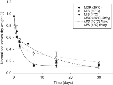
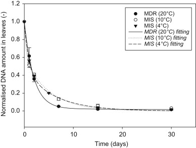
Fig. 1(left). Decreaseofleavesdrymatterwithtimeinsediments(normalizedbythe initial value).MDR(20℃) microcosmskeptindarkroomat20℃. MIS(4℃) microcosmskeptinthelakekeptat4℃. MIS(10℃) microcosmskeptinthelakeat 10℃.
Fig. 2(right). Decrease ofDNAweightwithtimeinleavesdrymatter(normalizedbythe initial value).MDR(20℃) microcosmskeptindarkroomat20℃. MIS(4℃) microcosmskeptinthelakekeptat4℃. MIS(10℃) microcosmskeptinthelakeat 10℃.
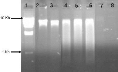
Fig. 3 Gel signature of DNA extracted from tomato leaves, water, and sediments at different time to estimate potential of DNA degradation. Lane 1: molecular weight STANDARD (Smart Ladder). Lane 2: DNA from fresh matter. Lane 3: DNA from dry matter not introduced in sediments. Lanes 4, 5, and 6: Tomato leaves DNA from microcosms MDR (20℃), MIS (10℃), and MIS (4℃), respectively, after 1 day of incubation time. Lanes 7and 8: DNA from lake water and sediments, respectively. 15µL of each extracted DNA were used.
Conclusion:
The results of this study suggest that (i) a two-compartment first-order model fitted both tomato dry matter and DNA content mass loss well, (ii) before release, an important quantity of DNA may be degraded inside plant tissues during decomposition in sediments and (iii) after release, DNA can both be rapidly adsorbed onto sediment particles and persist in water column as dissolved extracellular DNA, and (iv) water temperature and microbial activity were identified as the key factors influencing plant leaf decomposition and DNA stability. The present study has discussed the decomposition kinetics of plant material and the consequent DNA release in freshwater sediments. Both are important aspects for modelling DNA dispersion from plants into water column and sediments. The results of this study may help the field researcher understand what happens to plant DNA (transgenic or non-transgenic) when it is released and disperses into the aquatic environment. These are timely and relevant results in this field of research, with applications including the addition of transgenic DNA to aquatic environment DNA cycles. Further extension and application of this work should be performed with specific transgenes. The possibility of their horizontal transfer to sediment microorganisms is also worthy of investigation. The relative contribution of various plant communities (e.g. sediment bacteria or endo/exophytes) in facilitating extra versus intracellular plant DNA degradation also needs to be examined.
Yueting Pan