文献汇报:关于木质纤维素生物质转化的两种可视化研究
Article 1:拉曼光谱实现碱性预处理过程中木质纤维素的定量可视化
Title:Quantitative visualization of subcellular lignocellulose revealing the mechanism of alkali pretreatment to promote methane production of rice straw (https://doi.org/10.1186/s13068-020-1648-8)
Abstract:
Background: As a renewable carbon source, biomass energy not only helps in resolving the management problems of lignocellulosic wastes, but also helps to alleviate the global climate change by controlling environmental pollution raised by their generation on a large scale. However, the bottleneck problem of extensive production of biofuels lies in the flamentous crystal structure of cellulose and the embedded connection with lignin in biomass that leads to poor accessibility, weak degradation and digestion by microorganisms. Some pretreatment methods have shown signifcant improvement of methane yield and production rate, but the promotion mechanism has not been thoroughly studied. Revealing the temporal and spatial efects of pretreatment on lignocellulose will greatly help deepen our understanding of the optimization mechanism of pretreatment, and promote efcient utilization of lignocellulosic biomass. Here, we propose an approach for qualitative, quantitative, and location analysis of subcellular lignocellulosic changes induced by alkali treatment based on label-free Raman microspectroscopy combined with chemometrics.
Results: Firstly, the variations of rice straw induced by alkali treatment were characterized by the Raman spectra, and the Raman fngerprint characteristics for classifcation of rice straw were captured. Then, a label-free Raman chemical imaging strategy was executed to obtain subcellular distribution of the lignocellulose, in the strategy a serious interference of plant tissues’fuorescence background was efectively removed. Finally, the efects of alkali pretreatment on the subcellular spatial distribution of lignocellulose in diferent types of cells were discovered.
Conclusions: The results demonstrated the mechanism of alkali treatment that promotes methane production in rice straw through anaerobic digestion by means of a systemic study of the evidence from the macroscopic measurement and Raman microscopic quantitative and localization two-angle views. Raman chemical imaging combined with chemometrics could nondestructively realize qualitative, quantitative, and location analysis of the lignocellulose
of rice straw at a subcellular level in a label-free way, which was benefcial to optimize pretreatment for the improvement of biomass conversion efciency and promote extensive utilization of biofuel.
Keywords: Raman, Chemical imaging, Lignocellulosic biomass, Alkali pretreatment, Rice straw, Spectral unmixing
Result analysis:
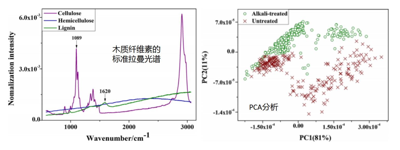
①下图为木质纤维素的标准拉曼光谱(580-3062cm-1)及拉曼光谱值的主成分分析(PCA)。结果表明纤维素有明显的峰(包括1089cm-1),几乎不受干扰,而木质素的峰出现在1620cm-1。此外,半纤维素和木质素受到荧光的干扰,尤其是半纤维素没有明显的峰。1620cm-1和1089cm-1这两个波段是未处理和碱处理稻草样品分类的谱带特征。此外,PCA结果显示拉曼光谱可以捕捉未经处理和碱处理的稻草样品的差异。
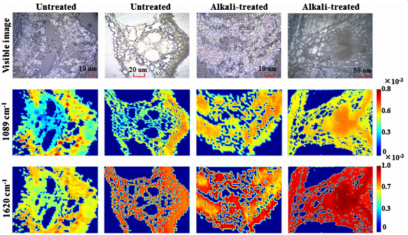
②下图为未处理和碱处理稻草横断面单一特征带的拉曼图像。拉曼谱带1089、1508、1620和1739cm-1都与木质纤维素有关。具体来说:1089cm-1的拉曼带可归因于半纤维素中的碳氧碳和碳碳环振动,以及纤维素中的碳氧碳拉伸,1620cm-1的峰与环共轭碳=碳链相关,1508cm-1附近的拉曼带强度与木质素中苯环的不对称拉伸振动有关,1739cm-1带区与半纤维素中的的C=O拉伸振动有关。下图中显示了1089cm-1和1620cm-1带区的单一特征带的拉曼图像。
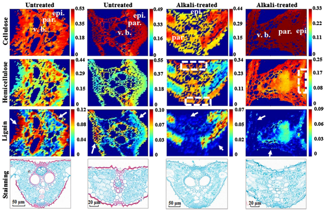
③基于木质纤维素标准品的参考光谱和单一特征带,用FCLS光谱解混,将其用作光谱全范围约束,得到显微拉曼图谱中的每个采样点解析的木质纤维素亚细胞分布的可靠图像。结构上:未处理的木质纤维素(UT)高度光滑、致密和均匀排列的多孔结构,轮廓清晰,细胞壁完整。碱处理后的木质纤维素(AT)预处理导致表面结构的严重损坏,形成新的图案,具有粗糙的表面、更多的裂纹和空隙以及大的表面积。同时,组织膨胀,微观结构破裂。空间分布上:UT组的纤维素分布均匀且密集,半纤维素集中在具有点状颗粒的薄壁组织中。木质素主要分布在表皮细胞、木质部和厚壁组织中。AT组显示,碱处理后,木质纤维素(纤维素、半纤维素和木质素)含量急剧下降,高度有序的空间分布也被破坏。值得注意的是,表皮细胞、木质部和厚壁组织中的大多数木质素被降解。碱处理导致木质素在横断面集中分布,较少靠近上表皮和下表皮。
Article 2:共聚焦激光扫描显微镜(CLSM)结合荧光标记实现木质纤维素在酶水解过程的结构变化和可视化
Title:Pretreatment of sweet sorghum straw and its enzymatic digestion: insight into the structural changes and visualization of hydrolysis process (https://doi.org/10.1186/s13068-019-1613-6)
Abstract:
Background: The efcient utilization of lignocellulosic biomass for biofuel production has received increasing attention. Previous studies have investigated the pretreatment process of biomass, but the detailed enzymatic hydrolysis process of pretreated biomass remains largely unclear. Thus, this study investigated the pretreatment efciency of dilute alkali, acid, hydrogen peroxide and its ultimate efects on enzymatic hydrolysis. Furthermore, to better understand the enzymatic digestion process of alkali-pretreated sweet sorghum straw (SSS), multimodal microscopy techniques were used to visualize the enzymatic hydrolysis process.
Result: After pretreatment with alkali, an enzymatic hydrolysis efciency of 86.44% was obtained, which increased by 99.54% compared to the untreated straw (43.23%). The FTIR, XRD and SEM characterization revealed a sequence of microstructural changes occurring in plant cell walls after pretreatment, including the destruction of lignin–polysaccharide interactions, the increase of porosity and crystallinity, and reduction of recalcitrance. During the course of hydrolysis, the cellulase dissolved the cell walls in the same manner and the digestion frstly occurred from the middle
of cell walls and then toward the cell wall corners. The CLSM coupled with fuorescent labeling demonstrated that the sclerenchyma cells and vascular bundles in natural SSS were highly lignifed, which caused the nonproductive bindings of cellulase on lignin. However, the efcient delignifcation signifcantly increased the accessibility and digestibility of cellulase to biomass, thereby improving the saccharifcation efciency.
Conclusion: This work will be helpful in investigating the biomass pretreatment and its structural characterization. In addition, the visualization results of the enzymatic hydrolysis process of pretreated lignocellulose could be used for guidance to explore the lignocellulosic biomass processing and large-scale biofuel production.
Keywords: Sweet sorghum straw, Pretreatment, Structural characterization, Enzymatic hydrolysis process, Visualization
Result analysis:
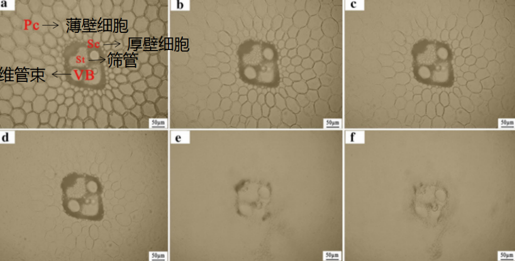
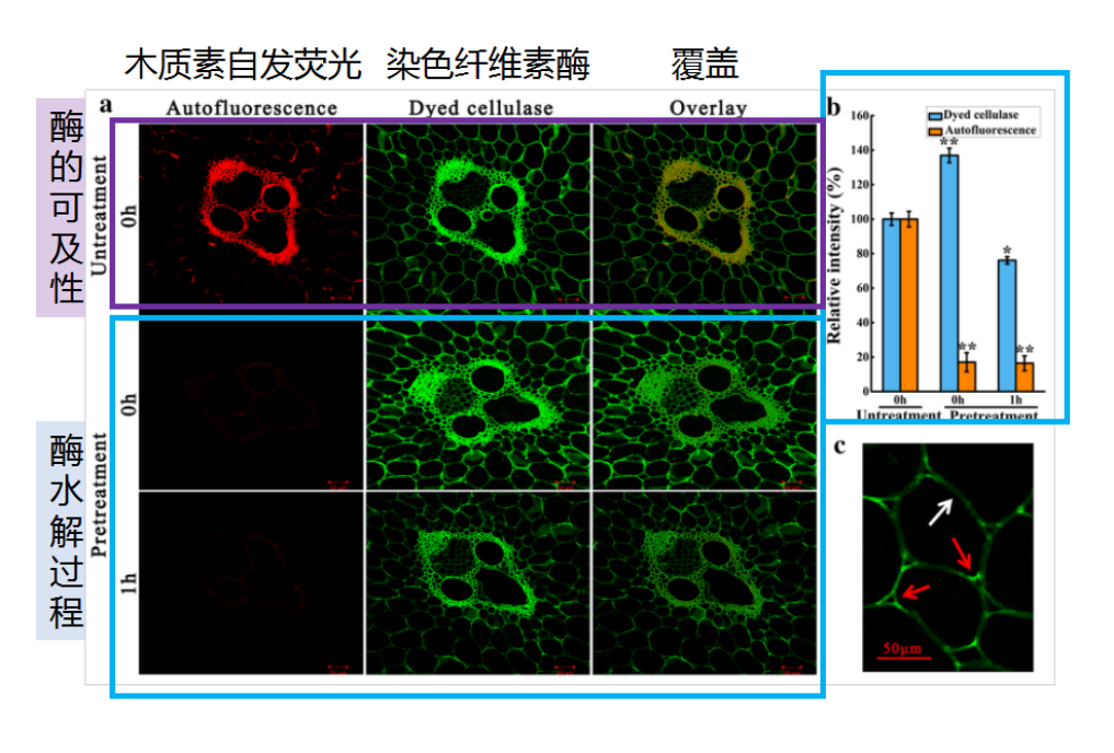
①下图为bright-feld光学显微镜的对照图像,观察对象为碱预处理后的甜高粱秸秆(横切面,20μm厚)在室温下的酶水解过程。以60分钟的时间间隔记录图像a-f 。结果表明远离维管束的薄壁细胞的水解速率比厚壁细胞和维管束的水解速率快得多。水解5小时后,在9 IU/g底物酶负载下观察到薄壁细胞完全降解维管束最终可以被降解。总消化率与预处理细胞壁中残余木质素含量呈负相关。
②下图为共聚焦激光扫描显微镜(CLSM)结合荧光标记,对高粱秸秆的切片进行观察,CLSM用于显示预处理生物质对纤维素酶的可及性和可消化性的空间变化,以及水解过程非均质组分含量的变化。结果显示,a:对于未经预处理的样品,标记的荧光非选择性地分布在薄壁细胞和高度木质化的维管束的横切面中,表明纤维素酶与木质素发生了非生产性结合。b:水解1小时后,染料标记的荧光强度下降,这归因于纤维素的部分消化导致纤维素中吸收的染色纤维素酶的分解。c:酶促消化首先从细胞壁的中部开始,然后向细胞壁的角落进行,这也得到CLSM结果的支持。
两种可视化成像的优势和比较
拉曼化学成像显微光谱:
可实现结构的无标记分析和量化,将振动光谱的化学特性与光学放大能力相结合,具有高空间分辨率和化学特异性等优点,且不受水的干扰。
共聚焦激光扫描显微镜(CLSM):
可以与荧光标记相结合,用于显示预处理生物质对纤维素酶的可及性和可消化性的空间变化,以及水解过程非均质组分含量的变化。
Contact: ZhouMin
Email:986624183@qq.com